Oral Presentation ANZBMS-MEPSA-ANZORS 2022
Anterior lift-off of implanted press-fit tibial tray during deep knee bend: a time-elapsed micro-CT and digital volume correlation analysis. (#59)
Achieving primary stability of press-fit tibial implants is important for their long-term success. Deep knee bending (DKB) exposes the implant to posterior loading that may jeopardise this stability1. Here, time-elapsed micro-CT imaging and digital volume correlation (DVC) is undertaken on a cadaveric human tibia implanted with a titanium tibial tray, while under a mechanical loading sequence replicating DKB, with the aim of quantifying the internal strain field under progressive load.
One cadaveric proximal tibia (male: 42yr, 67kg) was implanted with a titanium tibial tray. Step-wise uniaxial loads (0.0–3.5 x body weight (BW)) were applied through two contact points replicating the medial and lateral condyle of the femur on the tray during DKB. Micro-CT scans (46µm/pixel) were performed at each load step. Loaded scans were co-registered to the 0.0BW scan and DVC analysis performed (DaVis, LaVision). The minimum principal strains (Pmin) of subvolumes in direct contact with the tray, and then moving radially outwards, were extracted, and their 10th percentile analysed (Fig.1a). For five volumes of interest (VOIs; pegs: anterior-lateral (AL), anterior-medial (AM), posterior-lateral (PL), posterior-medial (PM) and Tray), the strain values were normalised to the final load step.
Increased compressive strains reflected the progressive compressive loads applied, while bone strain dissipated radially from the implant. Interestingly, at 1.5BW there was a reduction of Pmin across the anterior pegs (AL, AM) (Fig.1b), which coincided with an anterior lift-off of the tray, observed from the micro-CT images.
This ongoing study presents a novel method to analyse the internal strain of an implanted proximal tibia under progressive load, which will improve the understanding of primary implant stability.
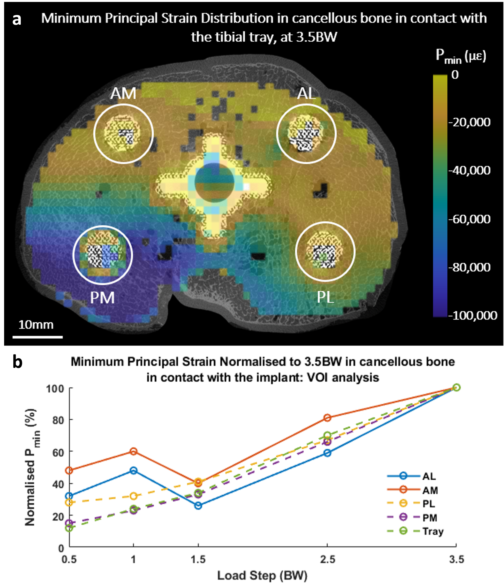
Fig.1a) Minimum principal strain Pmin (µε) distribution at 3.5BW; b) Normalised Pmin (%, normalised to the 3.5BW load step) in each volume of interest (VOI).
- 1.Taylor et al., J Orthop Res, 30:1362-1368, 2012